We use our brains to think every day, but the human brain is so complex, subtle, and mysterious that we still know very little about it. However, the development of brain imaging technology over the years has brought new advances to scientists, who study how the human brain works by using the device like kernel flow.
What is Kernel Flow?
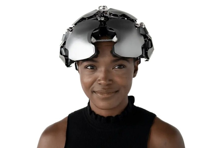
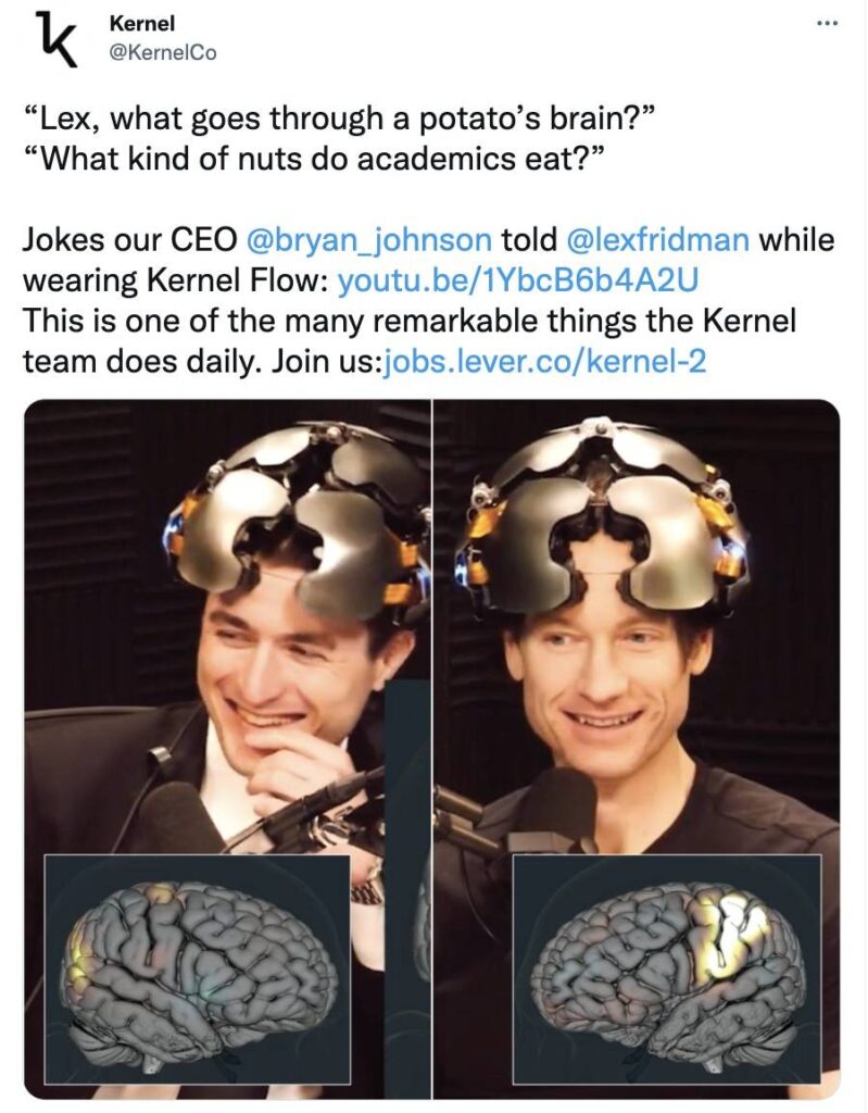
Researchers at Kernel, a neurotechnology startup in the United States, have developed a wearable device called Kernel Flow, which is a brain-reading helmet. This helmet device uses TD-fNIRS (Time Domain Functional Near Infrared Spectroscopy) technology to measure brain activity by recording local changes in oxygen content of blood.
Most non-invasive brain-scanning systems adopt fNIRS technology. It means that a beam of light in the near infrared spectrum is emitted to increase blood flow volume to the brain, in order to measure changes in light absorption by hematoglobulin in the brain’s blood-circulating system. As has been applying for a long time, TD-fNIR is considered as the gold standard for the invention of non-invasive optical imaging of brain devices, but problems such as high cost, complexity, large size, and poor sampling frequency limit its application.
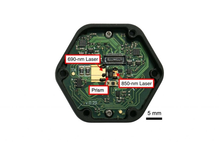
Kernel Flow consists of 52 modules distributed in four regions covering the frontal lobe, parietal lobe, temporal lobe and occipital cortex. Each module contains a detector and a laser source that emits laser light at two different wavelengths, 690nm and 850nm, and travels through the scalp to the brain.
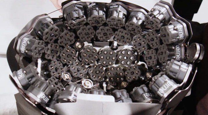
There are six detectors arranged in a hexagonal shape around the laser source, each at a distance of 10 mm from the light source. The detector picks up the reflected light, and records the arrival time of photon, detecting approximately more than one billion photons per second.
The detectors record the detected photon arrival times as a histogram at a sampling rate of 200Hz, with a sampling frequency of 7.1 Hz for the entire system. Designing for dealing with high photon count rates, these detectors are optimized for ToF measurements of diffuse reflectance tomography, with processing speeds in excess of 1 × 10⁹/sec.
Scientific test with Kernel Flow
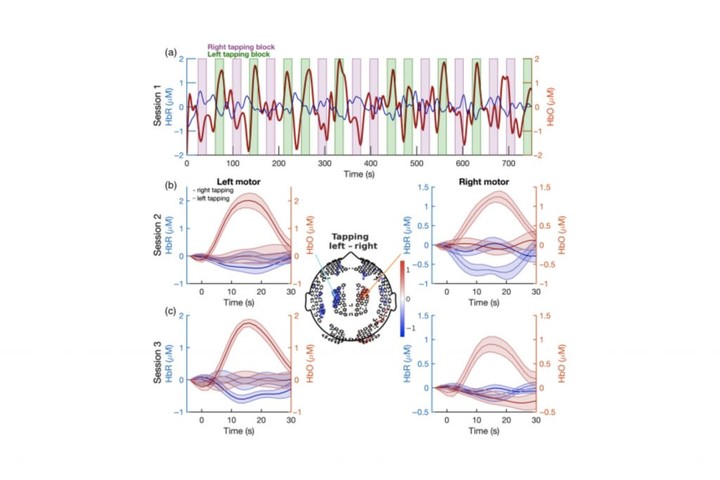
In a neuroscience test conducted by Kernel, two volunteers who participated showed significant hemodynamic changes in channels in the motor cortex during a finger-tapping task. In addition, Kernel Flow’s TD-fNIR system identified heartbeat oscillations on the forehead of one of the volunteers with the help of high sampling rates.
In addition to these tests, Kernel plans to use Flow to conduct different studies. For example, brain images are used to understand emotions, side effects of psychedelic drugs such as ketamine, and attention span and others. However, the Kernel researchers say the technology currently has certain limitations, such as hair texture and skin type that can affect the results of brain imaging.
The current kernel flow helmet weighs only 2.2 kilograms and is built with a small laser driver, custom integrated circuit and dedicated detector, already performs similarly to a benchtop system. While commercial viability has yet to improve, it may not be long before we can measure brain function in the same way that we measure heart rate.
Introduction to the principle of near infrared brain imaging technology
Due to the sensitivity to motion artifacts, electromagnetic interference, and the claustrophobic imaging process, existing brain functional imaging techniques such as functional magnetic resonance imaging (fMRI), are not suitable for children (especially infants), nor for natural research and application of high ecological validity scenarios. Functional near infrared spectroscopy (fNIRS) is considered to be a functional brain imaging technology that can meet the above requirements, and provides a new observation method for basic research and clinical applications of brain science.
fNIRS fundamentals
Based on the characteristics of strong penetration of near-infrared light, by placing different numbers of near-infrared light emitting probes and near-infrared light absorption probes on the surface of the volunteer’s head, then irradiating near-infrared light and measuring the absorption of near-infrared light by brain tissue with single or multiple channels.
While the light absorption changes, various indicators in the corresponding regions of the cerebral cortex are measured, including HbO2, Hb, total oxyhemoglobin (tHb), oxidized cytochrome oxidase (Cytox), brain Blood flow (CBF), cerebral blood volume (CBV), tissue oxygenation index (TOI), local oxygen saturation (ISO), etc. The changes of each index are reconstructed in two-dimensional or three-dimensional images, and the functional state of the cerebral cortex is reflected through the display of dynamic images.
Principle of fNIRS device
The optical tomography device such as the kernel flow helmet we talked before emits a near-infrared light source, which is transported to the measurement site through a flexible optical fiber, and receives the attenuated reflection signal from the tissue from the detector. When the distance between the source and detector is 2.0cm~3.0cm, the near infrared light can reach a suitable measurement depth, that is the cerebral cortex. The flexible optical fiber can meet the various postures and head positions of the volunteers, and the physical activity does not need to be strictly restricted. The measurement of fNIRS can be done in a daily life environment without the need for volunteers to apply sedatives, and it is also convenient and applicable to infants and children.
Based on the characteristics of high scattering and low absorption of human tissue, the detection system adopts a reflective detection method, the light source and the detector are located on the same side of the detected object. By simulating the transmission process of photons in the human brain by the Monte Carlo method, it is found that the propagation path of photons in the brain is banana-shaped, and when the distance between the light source and the detector exceeds 2.9cm, the photons can penetrate at least 1cm depth. Based on the anatomical structure of the brain, it can penetrate the outer tissues and reach the gray matter, so that it can carry information on the hemoglobin content of the gray matter. The farther the light source is from the detector, the greater the penetration depth and the greater the loss of light.
Beer-Lambert Law
There are abundant microvascular in the tissue, with diameter <1mm, including capillaries, micro -artery, micro -veins, etc. The red blood cells transported by microvascular are carried with the main absorption body of near infrared light in the tissue: hemoglobin. This kind of protein plays important role to transport oxygen. After passing through the micro blood vessels, nearly infrared light will weaken, the higher the concentration of hemoglobin, and the more obvious the light weakens. In the case of knowing strong incident light and strong output light, the concentration of hemoglobin can be estimated, this law is summarized to quantify the law of Beer-Lambert.
Therefore, by determining the intensity of scattered light in the brain activity area, the blood oxygen and blood capacity of the area can be inferred. In this way, the relative concentration changes of the oxygen concentration in the brain and deoxymal hemoglobin can be obtained during cognitive activities, so as to speculate the relationship between the brain and the brain areas related to cognitive activities.
Researchers can use functional near infrared spectrum technology (FNIRS) to obtain changes in blood oxygen levels when brain activity, and then study the neural mechanism of cognitive activities.
Enjoy this? Then read more guides about gigabit switches, network cards and wifi routers.